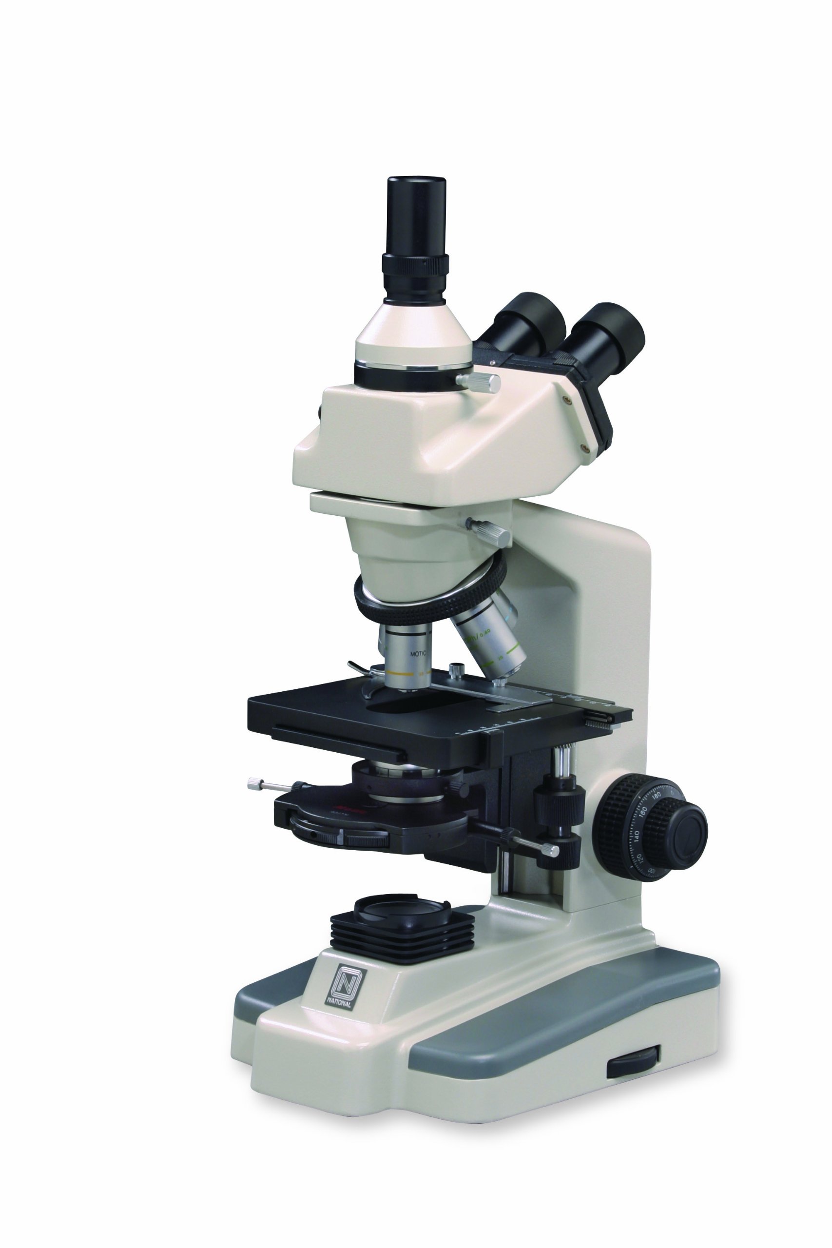
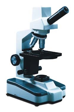
44 light microscope with labels
Compound Microscope Parts - Labeled Diagram and their Functions - Rs ... The eyepiece (or ocular lens) is the lens part at the top of a microscope that the viewer looks through. The standard eyepiece has a magnification of 10x. You may exchange with an optional eyepiece ranging from 5x - 30x. [In this figure] The structure inside an eyepiece. The current design of the eyepiece is no longer a single convex lens. Microscope Parts and Functions The specimen is placed on the glass and a cover slip is placed over the specimen. This allows the slide to be easily inserted or removed from the microscope. It also allows the specimen to be labeled, transported, and stored without damage. Stage: The flat platform where the slide is placed.
Compound Microscope Parts, Functions, and Labeled Diagram Abbe Condenser: This lens condenses the light from the base illumination and focuses it onto the stage. This piece of the compound microscope sits below the stage & typically acts as a structural support that connects the stage to arm or frame of the microscope. Coarse and fine adjustment controls: Adjusts the focus of the microscope.

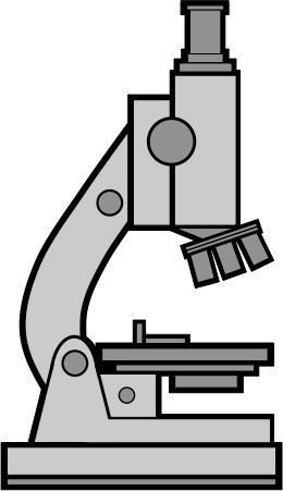
Light microscope with labels
› books › NBK26880Looking at the Structure of Cells in the Microscope ... Many light-microscope techniques are available for observing cells. Cells that have been fixed and stained can be studied in a conventional light microscope, while antibodies coupled to fluorescent dyes can be used to locate specific molecules in cells in a fluorescence microscope. Living cells can be seen with phase-contrast, differential ... Light Microscope- Definition, Principle, Types, Parts, Labeled Diagram ... A light microscope is a biology laboratory instrument or tool, that uses visible light to detect and magnify very small objects and enlarge them. They use lenses to focus light on the specimen, magnifying it thus producing an image. The specimen is normally placed close to the microscopic lens. Simple Microscope - Diagram (Parts labelled), Principle, Formula and Uses Optical microscope - These use transparent lenses and visible light to see objects to the order of one micrometer ( one millionth of one meter Charged Particle (ion and electron) microscope - These microscopes employ electrostatic or electromagnetic lens along with a beam of charged particles to focus on specimens. These microscopes can see objects as tiny as one nanometer ( one tenth billionth of one meter)
Light microscope with labels. Microscope Labeling - The Biology Corner Students label the parts of the microscope in this photo of a basic laboratory light microscope. Can be used for practice or as a quiz. Name_____ Microscope Labeling . Microscope Use: 15. When focusing a specimen, you should always start with the _____ objective. Required practical - using a light microscope - BBC Bitesize Care must be taken when handling coverslips and microscope slides. Drawing the image Record the microscope images using labelled diagrams or produce digital images. When first examining cells or... Labeling the Parts of the Microscope Labeling the Parts of the Microscope This activity has been designed for use in homes and schools. Each microscope layout (both blank and the version with answers) are available as PDF downloads. You can view a more in-depth review of each part of the microscope here. Download the Label the Parts of the Microscope PDF printable version here. › AmScope-LED-144W-ZK-AdjustableAmScope LED-144W-ZK White Adjustable 144 LED Ring Light ... Buy AmScope LED-144W-ZK White Adjustable 144 LED Ring Light Illuminator for Stereo Microscope ... DYMO Authentic LW Large Multi-Purpose Labels for LabelWriter Label ...
en.wikipedia.org › wiki › Electron_microscopeElectron microscope - Wikipedia An electron microscope is a microscope that uses a beam of accelerated electrons as a source of illumination. As the wavelength of an electron can be up to 100,000 times shorter than that of visible light photons, electron microscopes have a higher resolving power than light microscopes and can reveal the structure of smaller objects. Parts of the Microscope with Labeling (also Free Printouts) Parts of the Microscope with Labeling (also Free Printouts) A microscope is one of the invaluable tools in the laboratory setting. It is used to observe things that cannot be seen by the naked eye. Table of Contents 1. Eyepiece 2. Body tube/Head 3. Turret/Nose piece 4. Objective lenses 5. Knobs (fine and coarse) 6. Stage and stage clips 7. Aperture Parts of a microscope with functions and labeled diagram Microscope Definition. Microscopes are instruments that are used in science laboratories to visualize very minute objects such as cells, and microorganisms, giving a contrasting image that is magnified. Microscopes are made up of lenses for magnification, each with its own magnification powers. A Study of the Microscope and its Functions With a Labeled Diagram These labeled microscope diagrams and the functions of its various parts, attempt to simplify the microscope for you. However, as the saying goes, 'practice makes perfect', here is a blank compound microscope diagram and blank electron microscope diagram to label.
Αποστείρωση και εργαστηριακή διάγνωση - Αποτέλεσμα Google Books Nikolas Morein · Medical"Realistic modeling of the illumination point spread function in confocal scanning optical microscopy". J. Opt. Soc. Am. A. 27 (2): 295–302. Microscope, Microscope Parts, Labeled Diagram, and Functions Majority of high quality microscopes used in laboratory include an Abbe condenser with an iris diaphragm. When iris diaphragm is combined with Abbe condenser, it control both the quantity of light applied as well as focus on the specimen. Aperture: It is the hole in the stage through which the base (transmitted) light reaches the stage. Light microscopes - Cell structure - Edexcel - BBC Bitesize Microscopes are used to produce magnified images. There are two main types of microscope: light microscopes are used to study living cells and for regular use when relatively low magnification and... Light Microscopy - an overview | ScienceDirect Topics Terminal bars, as viewed by light microscopy, are sites of apparent attachment of epithelial cells that have been shown to be structures that are continuous around the circumference of the entire cell. Terminal bars occupy restricted regions of the cell located in the vicinity of its apex.
32 Compound Light Microscope With Label - Labels For You
Compound Light Microscopes | Products | Leica Microsystems Leica Microsystems products are developed with the user in mind. That means our light microscopes know what matters most: superlative image quality, ergonomic handling, fast results and cost efficiency. System solutions consisting of application-oriented software and hardware components make our instrument even more efficient.

Microscope Picture To Label - Micropedia
Label the microscope — Science Learning Hub All microscopes share features in common. In this interactive, you can label the different parts of a microscope. Use this with the Microscope parts activity to help students identify and label the main parts of a microscope and then describe their functions. Drag and drop the text labels onto the microscope diagram. If you want to redo an answer, click on the box and the answer will go back to the top so you can move it to another box.

microscopic organisms - nwnature.net
› products › microscopeMicroscope Objective Lens | Products | Leica Microsystems The objective lens is a critical part of the microscope optics. The microscope objective is positioned near the sample, specimen, or object being observed. It has a very important role in imaging, as it forms the first magnified image of the sample. The numerical aperture (NA) of the objective indicates its ability to gather light and largely determines the microscope’s resolution, the ...
Labeling A Compound Light Microscope - ClipArt Best
Labelled Diagram Of A Light Microscope - GlobalSpec Products/Services for Labelled Diagram Of A Light Microscope Microscopes - (706 companies) ...and transmission electron microscopes. Acoustic and ultrasonic microscopes use sound waves to create images of the sample. Compound microscopes use a single light path. These types of microscopes can have a single eyepiece (monocular) or a dual eyepiece...

Rens blog : Science, cells
rsscience.com › stereo-microscopeParts of Stereo Microscope (Dissecting microscope) – labeled ... A stereo microscope allows you to see the surface of specimens with a 3-dimensional view. Under a stereo microscope, you can see the metallic texture and colors of the mosquito’s compound eyes. In contrast, the light has to pass through the specimen to form the image under a compound microscope.
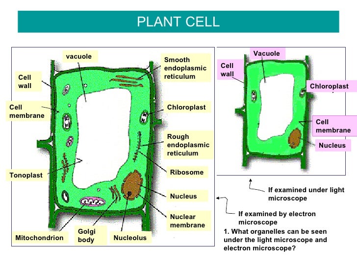
Bio F4 Cell Organel
Light Microscope Worksheet Live worksheets > English > Science > Lab equipment > Light Microscope Worksheet. Light Microscope Worksheet. Drag and drop worksheet on the parts of the microscope. ID: 1605909. Language: English. School subject: Science. Grade/level: Middle School. Age: 9-13. Main content: Lab equipment.

Label Compound Light Microscope - ClipArt Best
Microscope Labeling - The Biology Corner This simple worksheet pairs with a lesson on the light microscope, where beginning biology students learn the parts of the light microscope and the steps needed to focus a slide under high power. The labeling worksheet could be used as a quiz or as part of direct instruction where students label the microscope as you go over what each part is used for.
Drosophila sperm cells | Advanced Light Microscopy Facility
www2.nau.edu › lrm22 › lessonsMicroscope Notes - Northern Arizona University Microscope Drawings. When drawing what you see under the microscope, follow the format shown below. It is important to include a figure label and a subject title above the image. The species name (and common name if there is one) and the magnification at which you were viewing the object should be written below the image.
Search in gallery
Labeling the Parts of the Microscope | Microscope activity, Science ... Jan 13, 2016 - Free worksheets for labeling parts of the microscope including a worksheet that is blank and one with answers. ... Low power objective 4. Medium power objective 5. High power objective 6. Stage clips 7. Diaphragm 8. Light source 9. Eyepiece 10. Arm 11. Stage 12. Coarse adjustment knob 13. Fine adjustment knob 14. Base.
Labeling A Compound Light Microscope - ClipArt Best
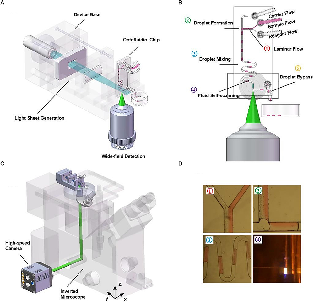
Light Sheet Microscopy | Science Lab | Leica Microsystems Light sheet microscopy - also referred to as single plane illumination microscopy or SPIM - is a gentle way of imaging sensitive samples or fast biological processes in vivo. The specimen is illuminated only in a single plane at a time and detected from the perpendicular direction. Since there is no out-of-focus excitation, phototoxic ...

Compound Light Microscope Labeled - Made By Creative Label
A quick guide to light microscopy in cell biology - PMC Light microscopy is a key tool in modern cell biology. Light microscopy has several features that make it ideally suited for imaging biology in living cells: the resolution is well-matched to the sizes of subcellular structures, a diverse range of available fluorescent probes makes it possible to mark proteins, organelles, and other structures for imaging, and the relatively nonperturbing ...

33 Label A Compound Light Microscope - Labels Database 2020
Labeling Microscope Worksheet | Teaching Resources docx, 300.56 KB. A straightforward worksheet in which students are required to identify the parts of a basic microscope. Tes classic free licence.

Histology Drawings: January 2014
› microscopy › intSmart Microscope for Lab Routine and Research - ZEISS Acquiring fluorescent images has never been so easy. Combine Axioscope 5 with the LED light source Colibri 3 and the sensitive, standalone microscope camera Axiocam 202 mono to have the perfect setup for easy multichannel fluorescence documentation. Switch effortlessly between the channels for UV, blue, green and red excitation.
33 Label All Indicated Parts Of The Microscope - Labels Database 2020
An Introduction to the Light Microscope, Light Microscopy Techniques ... Figure 1: Basic compound microscope: Light from a source is focused onto the sample (object) using a mirror and condenser lens. Light from the sample is collected by an objective, forming an intermediate image which is imaged again by the eyepiece and relayed to the eye, which sees a magnified image of the sample.
compound light microscope clipart 10 free Cliparts | Download images on Clipground 2021
Microscope Drawing And Label - Painting Valley Label The Microscope... 270x350 14 0 Compound Light Micro... 630x380 8 1 Labeling The Parts O... 525x450 7 0 Compound Microscope ... 413x424 6 0 Microscope - Microsc... 236x262 4 1 Label Microscope Dia... 459x457 4 0 Microscope Parts Dia... 576x400 3 0 Section Cells View A... 512x346 2 1 Exe - Microscope Dra... 933x1163 2 0 Drawing - Microscope...
Quia - Protist Vocabulary
Ιατρική Μικροβιολογία II: Αποστείρωση, Εργαστηριακή Διάγνωση ... Merim Kumars, Gerald Dunders, Nikolas Morein · MedicalDesign for a Fourier-transform holographic microscope. Proceedings of International Symposium on X-Ray Microscopy II, Springer Series in Optical Sciences.
Post a Comment for "44 light microscope with labels"